
Science and Technology
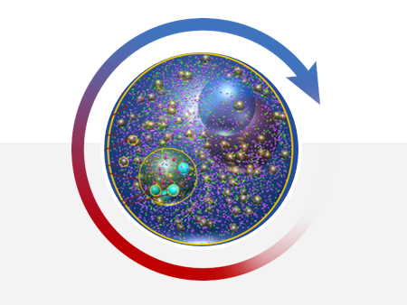
Hypothesis Generation
》Curated Knowledge-graph
》Network Al algorithms
》Data-mining integrations
》Virtual cell simulations
》Network Al algorithms
》Data-mining integrations
》Virtual cell simulations
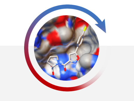
Chemistry ID & Optimization
》10 million compound library
》GPU-enabled docking tools
》Automated screening workflow
》GPU-enabled docking tools
》Automated screening workflow
Patient Selection
》50TB of patient data
》Feature-selection method
》Regression Al algorithms
》Classification Al
》Feature-selection method
》Regression Al algorithms
》Classification Al